
Top Level Name
⌊ Superfamily (core) Haloacid Dehalogenase
⌊ Subgroup C1.5: HAD, Beta-PGM, Phosphatase Like
Family known  |
|||||||
| Total |
100% |
<100% |
Family unknown |
||||
| Functional domains | 22318 | 0 | 812 | 21506 | |||
| UniProtKB | 3039 | 0 | 0 | 3039 | |||
| GI | 5264 | 0 | 0 | 5264 | |||
| Structures | 272 | ||||||
| Reactions | 0 | ||||||
| Functional domains of this subgroup were last updated on Nov. 22, 2017 | |||||||
| New functional domains were last added to this subgroup on Aug. 1, 2014 | |||||||
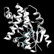
Structurally characterized enzymes in this subgroup have the C1 or C0 (only small inserts seen at either of the two points of cap insertion) cap type. Characterized functions included in this subgroup include 2-haloacid dehalogenase, beta-phosphoglucomutase, phosphonoacetaldehyde hydrolase, and phosphatases of various specificities.
Lahiri, S.D., et al.
Caught in the act: the structure of phosphorylated beta-phosphoglucomutase from Lactococcus lactis
▸ Abstract
Biochemistry 2002;41(26):8351-8359 | PubMed ID: 12081483
No notes.
Static File Downloads
| File Name | Description | Parameters | Stats |
|---|---|---|---|
| repnet.sg1129.th50.pE20.mek250.xgmml | Representative network: each node is a group of similar sequences | node similarity threshold = 50 max edge count = 250 min -log10 E = 20 |
size = 79M num_edges = 195537 num_nodes = 4983 |
| sfld_alignment_sg1129.msa | Annotated Sequence Alignment, Stockholm format | 57 sequences size: 33K |
| Subgroup ▸ Legend | T |
K |
C |
U |
S |
||
|---|---|---|---|---|---|---|---|
| C1.5: HAD, Beta-PGM, Phosphatase Like | 22318 | 812 | 0 | 21506 | 272 | ||
| ┗ 1: C1.5.1: Epoxide Hydrolase Phosphatase Like | 1561 | 7 | 0 | 1554 | 102 | ||
| ┗ epoxide hydrolase n-terminal phosphatase | 7 | 7 | 0 | 27 | |||
| ┗ 1: C1.5.2: MDP Like | 321 | 2 | 0 | 319 | 4 | ||
| ┗ mdp-1 | 2 | 2 | 0 | 2 | |||
| ┗ 1: C1.5.3: 5'-Nucleotidase Like | 630 | 0 | 0 | 630 | 15 | ||
| ┗ 1: C1.5.4: Enolase-phosphatase Like | 614 | 249 | 0 | 365 | 3 | ||
| ┗ enolase-phosphatase | 249 | 249 | 0 | 3 | |||
| ┗ 1: C1.5.5: Heptose Bisphosphate Phosphatase Like | 1994 | 3 | 0 | 1991 | 24 | ||
| ┗ histidinol-phosphatase | 3 | 3 | 0 | 0 | |||
| ┗ 1: C1.5.6: HAD, Beta-PGM, Phosphatase Like | 16451 | 551 | 0 | 15900 | 116 | ||
| ┗ 2-deoxyglucose-6-phosphatase | 7 | 7 | 0 | 0 | |||
| ┗ 2-haloacid dehalogenase | 145 | 145 | 0 | 12 | |||
| ┗ beta-phosphoglucomutase | 176 | 176 | 0 | 21 | |||
| ┗ glycerol-3-phosphate phosphatase | 5 | 5 | 0 | 1 | |||
| ┗ phosphoglycolate phosphatase | 75 | 75 | 0 | 0 | |||
| ┗ phosphonoacetaldehyde hydrolase | 143 | 143 | 0 | 8 |