Top Level Name

  ⌊ Superfamily (core) Haloacid Dehalogenase

    ⌊ Subgroup C1.5: HAD, Beta-PGM, Phosphatase Like

  C1.5.6: HAD, Beta-PGM, Phosphatase Like

     ⌊ Family beta-phosphoglucomutase

  ⌊ FunctionalDomain beta-phosphoglucomutase (ID 2262)

Superfamily Assignment Evidence Code(s) FSM PubMed:9649311
Family Assignment Evidence Code CFM PubMed:12081483
This entry was last updated onNov. 22, 2017

References to Other Databases

Genbank

SpeciesGIAccessionProteome
Lactococcus lactis Taxon ID: 1358 502661210 WP_012897250.1 (RefSeq) URP
Lactococcus lactis subsp. lactis Taxon ID: 1360 736044825 AJA56351.1 (Genbank) URP
Lactococcus lactis Taxon ID: 1358 691499754 KGF76471.1 (Genbank) URP
Show All

Uniprot

Protein NameAccessionEC Number Identifier
Beta-phosphoglucomutase P71447 5.4.2.6 PGMB_LACLA (Swiss-Prot)

Sequence

Length of Enzyme (full-length): 221 | Length of Functional Domain: 218

1       10        20        30        40        50        60

MFKAVLFDLDGVITDTAEYHFRAWKALAEEIGINGVDRQFNEQLKGVSREDSLQKILDLA
DKKVSAEEFKELAKRKNDNYVKMIQDVSPADVYPGILQLLKDLRSNKIKIALASASKNGP
FLLERMNLTGYFDAIADPAEVAASKPAPDIFIAAAHAVGVAPSESIGLEDSQAGIQAIKD
SGALPIGVGRPEDLGDDIVIVPDTSHYTLEFLKEVWLQ
KQK
This shows the full-length sequence of the enzyme. The region of the functional domain is highlighted in black letters, while the residual residues are shown in grey. The Superfamily domain, when present, is shown using underlining. In many cases the functional domain is the full-length sequence.
Conserved catalytic residues (as determined by automated alignment to family, subgroup, or superfamily HMMs) are shown with teal highlighting . Conserved catalytic residues which do not matched the Conserved Alignment Residue are shown with maroon highlighting . Information regarding their function can be found in the Conserved Residues section below.
FASTA formatted full-length sequence.
BLAST this sequence against SFLD.
Scan SFLD-HMMs with this sequence.

Conserved Residues

Superfamily CAR This EFD conserves 2/2 Superfamily-specific Conserved Alignment Residues.
Position Amino Acid Location Function Role Evidence Code Reference
8 Asp (D) side chain nucleophile: forms covalent intermediate covalent catalysis -- reactant ISS
114 Ser (S) side chain interacts with substrate/intermediate substrate binding -- binding ISS
Subgroup CAR This EFD conserves 2/2 Subgroup-specific Conserved Alignment Residues.
Position Amino Acid Location Function Role Evidence Code Reference
8 Asp (D) side chain Mg2+ ligand, nucleophile: attacks phosphate moiety of intermediate to form covalent intermediate covalent catalysis -- reactant,
metal ligand -- binding
ICS PubMed:12081483
145 Lys (K) side chain None -- ICS PubMed:10956028 PubMed:12081483
Family CAR This EFD conserves 8/8 Family-specific Conserved Alignment Residues.
Position Amino Acid Location Function Role Evidence Code Reference
8 Asp (D) side chain Mg2+ ligand, nucleophile: attacks phosphate moiety of intermediate to form covalent intermediate covalent catalysis -- reactant,
metal ligand -- binding
ICS PubMed:12081483
10 Asp (D) side chain Mg2+ ligand, nucleophile: attacks phosphate moiety of cofactor to form covalent intermediate covalent catalysis -- reactant,
metal ligand -- binding
ICS PubMed:12081483
16 Thr (T) side chain interacts with phosphate moiety of intermediate substrate binding -- binding ICS PubMed:12081483
45 Lys (K) side chain interacts with phosphate moiety of intermediate substrate binding -- binding ICS PubMed:12081483
114 Ser (S) side chain interacts with phosphate moiety of intermediate substrate binding -- binding ICS PubMed:12081483
145 Lys (K) side chain interacts with phosphate moiety of intermediate substrate binding -- binding ICS PubMed:12081483
169 Glu (E) side chain Mg2+ ligand metal ligand -- binding ICS PubMed:12081483
170 Asp (D) side chain Mg2+ ligand metal ligand -- binding ICS PubMed:12081483

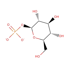
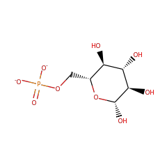
Catalyzed Reaction

beta-phosphoglucomutase

beta-D-glucose 1-phosphate(2-)
57684
beta-D-glucose 6-phosphate(2-)
58247

EC: 5.4.2.6 | IntEnz: 5.4.2.6 | Kegg: 5.4.2.6 | BioCyc: 5.4.2.6 | BRENDA: 5.4.2.6

Structures

PDB ID Title Molecule Name Number of
EFDs
Resolution (Å) Mutant? Het group Links
2WHE Structure Of Native Beta-Phosphoglucomutase In An Open Conformation Without Bound Ligands. Beta-Phosphoglucomutase 5 1.55 Magnesium Ion CSA • PDB • PDBSum
1Z4O Structure Of Beta-Phosphoglucomutase With Inhibitor Bound Alpha-Galactose 1-Phosphate Beta-Phosphoglucomutase 5 1.9 Magnesium Ion • 1-O-Phosphono-Alpha-D-Galactopyranose CSA • PDB • PDBSum
1Z4N Structure Of Beta-Phosphoglucomutase With Inhibitor Bound Alpha-Galactose 1-Phosphate Cocrystallized With Fluoride Beta-Phosphoglucomutase 5 1.97 Magnesium Ion • 1-O-Phosphono-Alpha-D-Galactopyranose CSA • PDB • PDBSum
2WFA Structure Of Beta-Phosphoglucomutase Inhibited With Beryllium Trifluoride, In An Open Conformation. Beta-Phosphoglucomutase 5 1.65 Beryllium Trifluoride Ion • Magnesium Ion CSA • PDB • PDBSum
1O08 Structure Of Pentavalent Phosphorous Intermediate Of An Enzyme Catalyzed Phosphoryl Transfer Reaction Observed On Cocrystallization With Glucose 1-Phosphate Beta-Phosphoglucomutase 5 1.2 Alpha-D-Glucose 1,6-Bisphosphate • Magnesium Ion CSA • PDB • PDBSum
2WF9 Structure Of Beta-Phosphoglucomutase Inhibited With Glucose-6-Phosphate, And Beryllium Trifluoride, Crystal Form 2 Beta-Phosphoglucomutase 5 1.4 Magnesium Ion
(4 more ⇓)
CSA • PDB • PDBSum
2WF8 Structure Of Beta-Phosphoglucomutase Inhibited With Glucose-6-Phosphate, Glucose-1-Phosphate And Beryllium Trifluoride Beta-Phosphoglucomutase 5 1.2 Beta-D-Glucose-6-Phosphate
(5 more ⇓)
CSA • PDB • PDBSum
1ZOL Native Beta-Pgm Beta-Phosphoglucomutase 5 1.9 Magnesium Ion CSA • PDB • PDBSum
1O03 Structure Of Pentavalent Phosphorous Intermediate Of An Enzyme Catalyzed Phosphoryl Transfer Reaction Observed On Cocrystallization With Glucose 6-Phosphate Beta-Phosphoglucomutase 5 1.4 Alpha-D-Glucose 1,6-Bisphosphate • Magnesium Ion CSA • PDB • PDBSum
2WF7 Structure Of Beta-Phosphoglucomutase Inhibited With Glucose-6-Phosphonate And Aluminium Tetrafluoride Beta-Phosphoglucomutase 5 1.05 6,7-Dideoxy-7-Phosphono-Beta-D-Gluco-Heptopyranose
(2 more ⇓)
CSA • PDB • PDBSum
2WF6 Structure Of Beta-Phosphoglucomutase Inhibited With Glucose-6-Phospahte And Aluminium Tetrafluoride Beta-Phosphoglucomutase 5 1.4 Magnesium Ion
(2 more ⇓)
CSA • PDB • PDBSum
2WF5 Structure Of Beta-Phosphoglucomutase Inhibited With Glucose-6-Phospahte And Trifluoromagnesate Beta-Phosphoglucomutase 5 1.3 Beta-D-Glucose-6-Phosphate
(2 more ⇓)
CSA • PDB • PDBSum
3ZI4 The Structure Of Beta-Phosphoglucomutase Inhibited With Glucose-6-Phospahte And Scandium Tetrafluoride Beta-Phosphoglucomutase 5 1.33 Scandium Tetraflouride
(2 more ⇓)
CSA • PDB • PDBSum
4C4T Structure Of Beta-Phosphoglucomutase In Complex With A Phosphonate Analogue Of Beta-Glucose-1-Phosphate And Aluminium Tetrafluoride Beta-Phosphoglucomutase 5 1.5 (S)-1-Beta-Phosphonofluoromethylene-1-Deoxy-D-Glucopyranose
(2 more ⇓)
CSA • PDB • PDBSum
4C4S Structure Of Beta-Phosphoglucomutase In Complex With An Alpha-Fluorophosphonate Analogue Of Beta-Glucose-1-Phosphate And Magnesium Trifluoride Beta-Phosphoglucomutase 5 1.5 (S)-1-Beta-Phosphonofluoromethylene-1-Deoxy-D-Glucopyranose
(2 more ⇓)
CSA • PDB • PDBSum
4C4R Structure Of Beta-Phosphoglucomutase In Complex With A Phosphonate Analogue Of Beta-Glucose-1-Phosphate And Magnesium Trifluoride Beta-Phosphoglucomutase 5 1.1 Beta-1 Phosphonomethylene-D-Glucopyranose
(2 more ⇓)
CSA • PDB • PDBSum
1LVH The Structure Of Phosphorylated Beta-Phosphoglucomutase From Lactoccocus Lactis To 2.3 Angstrom Resolution Beta-Phosphoglucomutase 5 2.3 Aspartyl Phosphate • Magnesium Ion CSA • PDB • PDBSum
3FM9 Analysis Of The Structural Determinants Underlying Discrimination Between Substrate And Solvent In Beta-Phosphoglucomutase Catalysis Beta-Phosphoglucomutase 5 2.7 Yes Magnesium Ion CSA • PDB • PDBSum
Percent identity and alignment length from the BLAST match of the Functional Domain sequence to the PDB sequence.
"Yes" indicates that a GI associated with this Functional Domain was mapped to the PDB ID via UniProtKB.

Curation History

Time Change Annotation Old Value New Value
Nov. 3, 2014, 9:26 a.m. update curation agent sbrown setDomainBoundaries.py
EC number assigned by UniProtKB accession ID.