Top Level Name

  ⌊ Superfamily (core) Haloacid Dehalogenase

    ⌊ Subgroup C2.B: Phosphomannomutase and Phosphatase Like

  C2.B.2: Mannosyl-3-phosphoglycerate Phosphatase Like

     ⌊ Family mannosyl-3-phosphoglycerate phosphatase

  ⌊ FunctionalDomain mannosyl-3-phosphoglycerate phosphatase (ID 2220)

Superfamily Assignment Evidence Code(s) ISS
Family Assignment Evidence Code CFM PubMed:11562374
This entry was last updated onNov. 22, 2017

References to Other Databases

Genbank

SpeciesGIAccessionProteome
Pyrococcus horikoshii Taxon ID: 53953 499187476 WP_010885016.1 (RefSeq)
Pyrococcus horikoshii OT3 Taxon ID: 70601 28380064 URP
Pyrococcus horikoshii OT3 Taxon ID: 70601 3257339 URP

Uniprot

Protein NameAccessionEC Number Identifier
Mannosyl-3-phosphoglycerate phosphatase {ECO:0000255|HAMAP-Rule:MF_00617, ECO:0000303|PubMed:11562374, ECO:0000303|PubMed:19018103} O58690 MPGP_PYRHO (Swiss-Prot)

Sequence

Length of Enzyme (full-length): 243 | Length of Functional Domain: 243

1       10        20        30        40        50        60

MIRLIFLDIDKTLIPGYEPDPAKPIIEELKDMGFEIIFNSSKTRAEQEYYRKELEVETPF
ISENGSAIFIPKGYFPFDVKGKEVGNYIVIELGIRVEKIREELKKLENIYGLKYYGNSTK
EEIEKFTGMPPELVPLAMEREYSETIFEWSRDGWEEVLVEGGFKVTMGSRFYTVHGNSDK
GKAAKILLDFYKRLGQIESYAVGDSYNDFPMFEVVDKAFIVGSLKHKKAQNVSSIIDVLE
VIK
This shows the full-length sequence of the enzyme. The region of the functional domain is highlighted in black letters, while the residual residues are shown in grey. The Superfamily domain, when present, is shown using underlining. In many cases the functional domain is the full-length sequence.
Conserved catalytic residues (as determined by automated alignment to family, subgroup, or superfamily HMMs) are shown with teal highlighting . Conserved catalytic residues which do not matched the Conserved Alignment Residue are shown with maroon highlighting . Information regarding their function can be found in the Conserved Residues section below.
FASTA formatted full-length sequence.
BLAST this sequence against SFLD.
Scan SFLD-HMMs with this sequence.

Conserved Residues

Superfamily CAR This EFD conserves 1/2 Superfamily-specific Conserved Alignment Residues.
Position Amino Acid Location Function Role Evidence Code Reference
40 Ser (S) side chain interacts with substrate/intermediate substrate binding -- binding ISS
Subgroup CAR This EFD conserves 7/7 Subgroup-specific Conserved Alignment Residues.
Position Amino Acid Location Function Role Evidence Code Reference
8 Asp (D) side chain Mg2+ ligand, nucleophile: attacks phosphate moiety of substrate to form covalent intermediate covalent catalysis -- reactant,
metal ligand -- binding
ICS PubMed:21961705
10 Asp (D) side chain general acid proton relay -- reactant ICS PubMed:21961705
40 Ser (S) side chain None -- ISS PubMed:21961705
42 Lys (K) side chain Positions general acid/base Asp steric role -- spectator ICS PubMed:21961705
180 Lys (K) side chain None -- ISS PubMed:21961705
204 Asp (D) side chain Mg2+ ligand metal ligand -- binding ICS PubMed:21961705
208 Asp (D) side chain None -- ISS PubMed:21961705
Family CAR This EFD conserves 14/14 Family-specific Conserved Alignment Residues.
Position Amino Acid Location Function Role Evidence Code Reference
8 Asp (D) side chain Mg2+ ligand; nucleophile: attacks phosphate moiety of substrate to form covalent intermediate covalent catalysis -- reactant,
metal ligand -- binding
ICS PubMed:21961705
10 Asp (D) side chain general acid: donates proton to leaving group proton relay -- reactant ICS PubMed:21961705
40 Ser (S) side chain None -- ISS PubMed:21961705
42 Lys (K) side chain Positions Asp general acid steric role -- spectator ICS PubMed:21961705
46 Glu (E) side chain Binds substrate substrate binding -- binding ICS PubMed:21961705
115 Tyr (Y) side chain Assists in stereochemical discrimination of mannosyl group from other sugar compounds steric role -- spectator ICS PubMed:21961705
127 Thr (T) side chain Forms hydrogen bonding network that helps fix mannosyl moiety in active site substrate binding -- binding ICS PubMed:21961705
140 Arg (R) side chain Binds C3(OH) hydroxyl of mannosyl moiety substrate binding -- binding ICS PubMed:21961705
145 Thr (T) side chain Assists in stereochemical discrimination of mannosyl group from other sugar compounds steric role -- spectator ICS PubMed:21961705
168 Gly (G) side chain Mg2+ ligand (via water); Stabilizes conformational change of C23O13(H) group, allowing nucleophilic attack on phosphate by activated water electrostatic stabiliser -- spectator,
metal ligand -- binding
ICS PubMed:21961705
170 Arg (R) side chain Helps close cap, fixing substrate in active site steric role -- spectator ICS PubMed:21961705
180 Lys (K) side chain None -- ISS PubMed:21961705
204 Asp (D) side chain Mg2+ ligand metal ligand -- binding ICS PubMed:21961705
208 Asp (D) side chain None -- ISS PubMed:21961705

Catalyzed Reaction

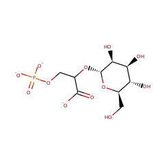
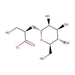
mannosyl-3-phosphoglycerate phosphatase

+ +
2-(alpha-D-mannosyl)-3-phosphonatoglycerate(3-)
57744
water
15377
2-(alpha-D-mannosyl)-D-glycerate
57541
hydrogenphosphate
43474

EC: | IntEnz: | Kegg: | BioCyc: | BRENDA: |

Structures

PDB ID Title Molecule Name Number of
EFDs
Resolution (Å) Mutant? Het group Links
1WZC Crystal Structure Of Pyrococcus Horikoshii Mannosyl-3-Phosphoglycerate Phosphatase Complexed With Mg2+ And Phosphate Mannosyl-3-Phosphoglycerate Phosphatase 3 1.9 Yes Magnesium Ion • Phosphate Ion CSA • PDB • PDBSum
2ZOS Crystal Structure Of Mannosyl-3-Phosphoglycerate Phosphatase From Pyrococcus Horikoshii Mannosyl-3-Phosphoglycerate Phosphatase 3 1.7 Yes Selenomethionine CSA • PDB • PDBSum
Percent identity and alignment length from the BLAST match of the Functional Domain sequence to the PDB sequence.
"Yes" indicates that a GI associated with this Functional Domain was mapped to the PDB ID via UniProtKB.

Curation History

Time Change Annotation Old Value New Value
Nov. 3, 2014, 9:26 a.m. update curation agent sbrown setDomainBoundaries.py
EC number assigned by UniProtKB accession ID.