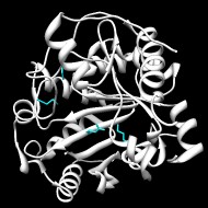
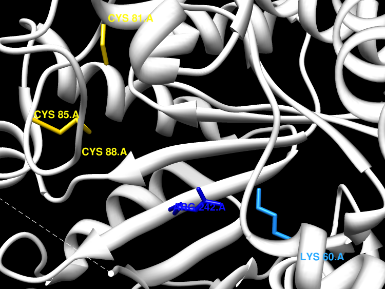
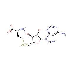
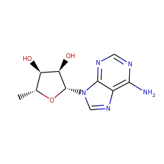
Wye bases are tricyclic bases that are found in archaeal and eukaryotic tRNAs. The most modified wye base, wybutosine, which appears at position 37 (the 30-adjacent position to the anti-codon), is known to be important for translational reading-frame maintenance. Saccharomyces cerevisiae TYW1 catalyses the tri-ring-formation step in wye-base biosynthesis, with the substrate tRNA bearing N1-methylated G37. Binds one [4Fe-4S] cluster and one [2Fe-2S cluster]. The 4Fe-4S cluster is coordinated with 3 cysteines and an exchangeable S-adenosyl-L-methionine. The two carbon source required for this reaction has recently been identified as pyruvate. It is thought that this reaction might proceed via a Schiff base mechanism. The exact role of the SAM molecule has yet to be determined, although it is shown here as acting in a stoichiometric manner, it could equally be catalytic in nature (and used to quench the .CO2 radical formed during the course of the proposed mechanism).
Goto-Ito S, Ishii R, Ito T, Shibata R, Fusatomi E, Sekine SI, Bessho Y, Yokoyama S
Structure of an archaeal TYW1, the enzyme catalyzing the second step of wye-base biosynthesis
▸ Abstract
Wye bases are tricyclic bases that are found in archaeal and eukaryotic tRNAs. The most modified wye base, wybutosine, which appears at position 37 (the 3'-adjacent position to the anticodon), is known to be important for translational reading-frame maintenance. Saccharomyces cerevisiae TYW1 catalyzes the tri-ring-formation step in wye-base biosynthesis, with the substrate tRNA bearing N(1)-methylated G37. Here, the crystal structure of the archaeal TYW1 homologue from Pyrococcus horikoshii is reported at 2.2 A resolution. The amino-acid sequence of P. horikoshii TYW1 suggested that it is a radical-AdoMet enzyme and the tertiary structure of P. horikoshii TYW1 indeed shares the modified TIM-barrel structure found in other radical-AdoMet enzymes. Radical-AdoMet enzymes generally contain one or two iron-sulfur (FeS) clusters. The tertiary structure of P. horikoshii TYW1 revealed two FeS cluster sites, each containing three cysteine residues. One FeS cluster site was expected from the amino-acid sequence and the other involves cysteine residues that are dispersed throughout the sequence. The existence of two FeS clusters was confirmed from the anomalous Fourier electron-density map. By superposing the P. horikoshii TYW1 tertiary structure on those of other radical-AdoMet enzymes, the AdoMet molecule, which is necessary for the reactions of radical-AdoMet enzymes, was modelled in P. horikoshii TYW1. Surface plots of conservation rates and electrostatic potentials revealed the highly conserved and positively charged active-site hollow. On the basis of the surface properties, a docking model of P. horikoshii TYW1, the tRNA, the FeS clusters and the AdoMet molecule was constructed, with the nucleoside at position 37 of tRNA flipped out from the canonical tRNA structure.
Yang Z, Shipman L, Zhang M, Anton BP, Roberts RJ, Cheng X
Structural characterization and comparative phylogenetic analysis of Escherichia coli HemK, a protein (N5)-glutamine methyltransferase
▸ Abstract
Protein glutamine methylation at GGQ sites of protein chain release factors plays a pivotal role in the termination of translation. We report here the crystal structure of the Escherichia coli HemK protein (N5)-glutamine methyltransferase (MTase) in a binary complex with the methyl-donor product S-adenosyl-L-homocysteine (AdoHcy). HemK contains two domains: a putative substrate binding domain at the N terminus consisting of a five helix bundle and a seven-stranded catalytic domain at the C terminus that harbors the binding site for AdoHcy. The two domains are linked by a beta-hairpin. Structure-guided sequence analysis of the HemK family revealed 11 invariant residues functioning in methyl-donor binding and catalysis of methyl transfer. The putative substrate-binding domains of HemK from E.coli and Thermotoga maritima are structurally similar, despite the fact that they share very little sequence similarity. When the two proteins are aligned structurally, the helical N-terminal domain is subject to approximately 10 degrees of hinge movement relative to the C-terminal domain. The apparent hinge mobility of the two domains may reflect functional importance during the reaction cycle. Comparative phylogenetic analysis of the hemK gene and its frequent neighbor gene, prfA, which encodes a major substrate, provides evidence for several examples of lateral gene transfer.
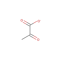
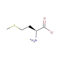
Pyruvate is the source of the two carbons that are required for formation of the imidazoline ring of 4-demethylwyosine
▸ Abstract
TYW1 catalyzes the condensation of N-methylguanosine with two carbon atoms from an unknown second substrate to form 4-demethylwyosine, which is a common intermediate in the biosynthesis of all of the hypermodified RNA bases related to wybutosine found in eukaryal and archaeal tRNA(Phe). Of the potential substrates examined, only incubation with pyruvate resulted in formation of 4-demethylwyosine. Moreover, incubation with C1, C2, C3, or C1,2,3-(13)C-labeled pyruvate showed that C2 and C3 are incorporated while C1 is not. The mechanistic implications of these results are discussed in the context of the structure of TYW1.
Mechanistic Studies of the Radical S-Adenosyl-L-methionine Enzyme 4-Demethylwyosine Synthase Reveal the Site of Hydrogen Atom Abstraction
▸ Abstract
TYW1 catalyzes the formation of 4-demethylwyosine via the condensation of N-methylguanosine (m¹G) with carbons 2 and 3 of pyruvate. In this study, labeled transfer ribonucleic acid (tRNA) and pyruvate were utilized to determine the site of hydrogen atom abstraction and regiochemistry of the pyruvate addition. tRNA containing a ²H-labeled m¹G methyl group was used to identify the methyl group of m¹G as the site of hydrogen atom abstraction by 5'-deoxyadenosyl radical. [2-¹³C₁-3,3,3-²H₃]Pyruvate was used to demonstrate retention of all the pyruvate protons, indicating that C2 of pyruvate forms the bridging carbon of the imidazoline ring and C3 the methyl.