Top Level Name

  ⌊ Superfamily (core) Enolase

    ⌊ Subgroup galactarate dehydratase

     ⌊ Family galactarate dehydratase 2

  ⌊ FunctionalDomain galactarate dehydratase (ID 366)

Superfamily Assignment Evidence Code(s) ISS
Family Assignment Evidence Code CFM PubMed:19883118
This entry was last updated onNov. 21, 2017

References to Other Databases

Genbank

SpeciesGIAccessionProteome
Oceanobacillus iheyensis Taxon ID: 182710 499379662 WP_011067240.1 (RefSeq)
Oceanobacillus iheyensis HTE831 Taxon ID: 221109 224510961 URP
Oceanobacillus iheyensis HTE831 Taxon ID: 221109 222447155 URP
Show All

Uniprot

Protein NameAccessionEC Number Identifier
Galactarate dehydratase (D-threo-forming) Q8EMJ9 GALRD_OCEIH (Swiss-Prot)

Sequence

Length of Enzyme (full-length): 391 | Length of Functional Domain: 391

1       10        20        30        40        50        60

MKITDLELHAVGIPRHTGFVNKHVIVKIHTDEGLTGIGEMSDFSHLPLYSVDLHDLKQGL
LSILLGQNPFDLMKINKELTDNFPETMYYYEKGSFIRNGIDNALHDLCAKYLDISVSDFL
GGRVKEKIKVCYPIFRHRFSEEVESNLDVVRQKLEQGFDVFRLYVGKNLDADEEFLSRVK
EEFGSRVRIKSYDFSHLLNWKDAHRAIKRLTKYDLGLEMIESPAPRNDFDGLYQLRLKTD
YPISEHVWSFKQQQEMIKKDAIDIFNISPVFIGGLTSAKKAAYAAEVASKDVVLGTTQEL
SVGTAAMAHLGCSLTNINHTSDPTGPELYVGDVVKNRVTYKDGYLYAPDRSVKGLGIELD
ESLLAKYQVPDLSWDNVTVHQLQDRTADTKS
This shows the full-length sequence of the enzyme. The region of the functional domain is highlighted in black letters, while the residual residues are shown in grey. The Superfamily domain, when present, is shown using underlining. In many cases the functional domain is the full-length sequence.
Conserved catalytic residues (as determined by automated alignment to family, subgroup, or superfamily HMMs) are shown with teal highlighting . Conserved catalytic residues which do not matched the Conserved Alignment Residue are shown with maroon highlighting . Information regarding their function can be found in the Conserved Residues section below.
FASTA formatted full-length sequence.
BLAST this sequence against SFLD.
Scan SFLD-HMMs with this sequence.

Conserved Residues

Superfamily CAR This EFD conserves 3/3 Superfamily-specific Conserved Alignment Residues.
Position Amino Acid Location Function Role Evidence Code Reference
193 Asp (D) side chain metal binding ligand metal ligand -- binding ISS PubMed:8987982
221 Glu (E) side chain metal binding ligand metal ligand -- binding ISS PubMed:8987982
245 Glu (E) side chain metal binding ligand metal ligand -- binding ISS PubMed:8987982
Subgroup CAR This EFD conserves 9/9 Subgroup-specific Conserved Alignment Residues.
Position Amino Acid Location Function Role Evidence Code Reference
42 Asp (D) side chain metal binding ligand (second metal) metal ligand -- binding ICS PubMed:19883118
45 His (H) side chain metal binding ligand (second metal) metal ligand -- binding ICS PubMed:19883118
90 Tyr (Y) side chain facilitates departure of beta-hydroxide leaving group; acid catalyst for ketonization of enol intermediate increase electrophilicity -- spectator,
proton relay -- reactant
ICS PubMed:19883118
162 Arg (R) side chain controls pKa of Tyr perturbates pKa -- spectator ICS PubMed:19883118
164 Tyr (Y) side chain abstracts proton from C2 of galactarate proton relay -- reactant ICS PubMed:19883118
193 Asp (D) side chain metal binding ligand metal ligand -- binding ICS PubMed:19883118
221 Glu (E) side chain metal binding ligand metal ligand -- binding ICS PubMed:19883118
246 His (H) side chain metal binding ligand metal ligand -- binding ICS PubMed:19883118
297 Thr (T) side chain metal binding ligand (second metal) metal ligand -- binding ICS PubMed:19883118
Family CAR This EFD conserves 9/9 Family-specific Conserved Alignment Residues.
Position Amino Acid Location Function Role Evidence Code Reference
42 Asp (D) side chain metal binding ligand (second metal) metal ligand -- binding ICS PubMed:19883118
45 His (H) side chain metal binding ligand (second metal) metal ligand -- binding ICS PubMed:19883118
90 Tyr (Y) side chain facilitates departure of beta-hydroxide leaving group; acid catalyst for ketonization of enol intermediate increase electrophilicity -- spectator,
proton relay -- reactant
ICS PubMed:19883118
162 Arg (R) side chain controls pKa of Tyr perturbates pKa -- spectator ICS PubMed:19883118
164 Tyr (Y) side chain abstracts proton from C2 of galactarate proton relay -- reactant ICS PubMed:19883118
193 Asp (D) side chain metal binding ligand metal ligand -- binding ICS PubMed:19883118
221 Glu (E) side chain metal binding ligand metal ligand -- binding ICS PubMed:19883118
246 His (H) side chain metal binding ligand metal ligand -- binding ICS PubMed:19883118
297 Thr (T) side chain metal binding ligand (second metal) metal ligand -- binding ICS PubMed:19883118

Catalyzed Reaction

galactarate dehydratase 2/3

+
galactarate(2-)
16537
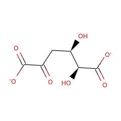
3-deoxy-D-threo-hex-2-ulosarate(2-)
78267
water
15377

EC: 4.2.1.- | IntEnz: 4.2.1.- | Kegg: 4.2.1.- | BioCyc: 4.2.1.- | BRENDA: 4.2.1.- |

Structures

PDB ID Title Molecule Name Number of
EFDs
Resolution (Å) Mutant? Het group Links
3GD6 Crystal Structure Of Divergent Enolase From Oceanobacillus Iheyensis Complexed With Phosphate Muconate Cycloisomerase 4 1.6 Phosphate Ion CSA • PDB • PDBSum
3ES8 Crystal Structure Of Divergent Enolase From Oceanobacillus Iheyensis Complexed With Mg And L-Malate. Muconate Cycloisomerase 4 2.2 (2S)-2-Hydroxybutanedioic Acid • Magnesium Ion CSA • PDB • PDBSum
2OQY The Crystal Structure Of Muconate Cycloisomerase From Oceanobacillus Iheyensis Muconate Cycloisomerase 4 2.0 Magnesium Ion CSA • PDB • PDBSum
3ES7 Crystal Structure Of Divergent Enolase From Oceanobacillus Iheyensis Complexed With Mg And L-Malate. Muconate Cycloisomerase 4 1.9 (2S)-2-Hydroxybutanedioic Acid • Magnesium Ion CSA • PDB • PDBSum
3FYY Crystal Structure Of Divergent Enolase From Oceanobacillus Iheyensis Complexed With Mg Muconate Cycloisomerase 4 1.8 Magnesium Ion CSA • PDB • PDBSum
3HPF Crystal Structure Of The Mutant Y90F Of Divergent Galactarate Dehydratase From Oceanobacillus Iheyensis Complexed With Mg And Galactarate Muconate Cycloisomerase 4 1.8 Yes D-Galactaric Acid • Magnesium Ion CSA • PDB • PDBSum
Percent identity and alignment length from the BLAST match of the Functional Domain sequence to the PDB sequence.
"Yes" indicates that a GI associated with this Functional Domain was mapped to the PDB ID via UniProtKB.

Identical Sequences with Suspect Annotations in Other Databases (Schnoes et.al. 2009)

Genbank

Source DB Accession Species Source Annotation Misannot. Evid. Code Source Date
NR 22778530 Oceanobacillus iheyensis HTE831 muconate cycloisomerase SFA Feb. 17, 2006
NR 23100298 Oceanobacillus iheyensis HTE831 muconate cycloisomerase SFA Feb. 17, 2006
TrEMBL Q8EMJ9_OCEIH Oceanobacillus iheyensis Muconate cycloisomerase SFA Feb. 17, 2006

Curation History

Time Change Annotation Old Value New Value
Nov. 3, 2014, 2:33 a.m. update curation agent sbrown setDomainBoundaries.py
EC number assigned by UniProtKB accession ID.