Top Level Name
⌊ Superfamily (core) Haloacid Dehalogenase
⌊ Subgroup C1.6: Phosphoserine Phosphatase Like
⌊ FunctionalDomain C1.6: Phosphoserine Phosphatase Like (ID 274546)
No Notes.
| Superfamily Assignment Evidence Code(s) | ISS |
| This entry was last updated on | Nov. 22, 2017 |
References to Other Databases
Genbank
| Species | GI | Accession | Proteome |
|---|---|---|---|
| Bacillus subtilis subsp. subtilis str. 168 Taxon ID: 224308 | 16078424 | NP_389243.1 (RefSeq) | PRP URP |
| Bacillus subtilis Taxon ID: 1423 | 489338546 | WP_003245748.1 (RefSeq) | URP |
| Bacillus subtilis Taxon ID: 1423 | 846136035 | AKN13482.1 (Genbank) | URP |
| Bacillus subtilis KCTC 1028 Taxon ID: 1136873 | 807073706 | AKC46912.1 (Genbank) | URP |
| Bacillus subtilis Taxon ID: 1423 | 760456530 | KIX82605.1 (Genbank) | URP |
| Bacillus subtilis subsp. subtilis Taxon ID: 135461 | 749185327 | AJE94037.1 (Genbank) | URP |
| Bacillus subtilis Taxon ID: 1423 | 728888573 | AIY96967.1 (Genbank) | URP |
| Bacillus subtilis subsp. subtilis str. 168 Taxon ID: 224308 | 728884262 | AIY92657.1 (Genbank) | PRP URP |
| Bacillus subtilis subsp. subtilis Taxon ID: 135461 | 672774597 | KFH34172.1 (Genbank) | URP |
| Bacillus subtilis subsp. subtilis Taxon ID: 135461 | 672773700 | KFH33276.1 (Genbank) | URP |
| Bacillus subtilis subsp. subtilis str. AG1839 Taxon ID: 1221328 | 649015076 | AIC43998.1 (Genbank) | URP |
| Bacillus subtilis subsp. subtilis str. JH642 substr. AG174 Taxon ID: 1232554 | 649010712 | AIC39766.1 (Genbank) | URP |
| Bacillus subtilis QH-1 Taxon ID: 1437006 | 588500428 | EXF52840.1 (Genbank) | URP |
| Bacillus subtilis subsp. subtilis 6051-HGW Taxon ID: 1147161 | 459389140 | AGG60726.1 (Genbank) | URP |
| Bacillus subtilis MB73/2 Taxon ID: 1267547 | 452117149 | EME07544.1 (Genbank) | URP |
| Bacillus subtilis QB928 Taxon ID: 1220533 | 402480780 | AFQ57289.1 (Genbank) | URP |
| Bacillus sp. Taxon ID: 1409 | 724427020 | URP | |
| Bacillus subtilis Taxon ID: 1423 | 723797826 | URP | |
| Bacillus subtilis BEST7003 Taxon ID: 1204342 | 407964336 | URP | |
| Bacillus subtilis BEST7613 Taxon ID: 1204343 | 407958758 | URP | |
| Bacillus subtilis subsp. subtilis str. 168 Taxon ID: 224308 | 41017265 | PRP URP | |
| Bacillus subtilis subsp. subtilis str. 168 Taxon ID: 224308 | 2633731 | PRP URP | |
| obsolete GIs = 433620000, 452914729, 221322747, 221318474, 221313552, 221309224, 670920356, 470161534, 402775597 | |||
| Show All | |||
Uniprot
| Protein Name | Accession | EC Number
 |
Identifier |
|---|---|---|---|
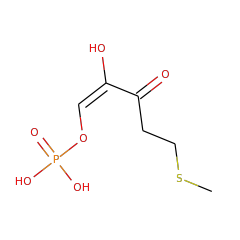
| 2-hydroxy-3-keto-5-methylthiopentenyl-1-phosphate phosphatase | O31667 | 3.1.3.87 | MTNX_BACSU (Swiss-Prot) |
Length of Enzyme (full-length): 235 | Length of Functional Domain: 223
MTTRKPFIICDFDGTITMNDNIINIMKTFAPPEWMALKDGVLSKTLSIKEGVGRMFGLLP
SSLKEEITSFVLEDAKIREGFREFVAFINEHEIPFYVISGGMDFFVYPLLEGIVEKDRIY
CNHASFDNDYIHIDWPHSCKGTCSNQCGCCKPSVIHELSEPNQYIIMIGDSVTDVEAAKL
SDLCFARDYLLNECREQNLNHLPYQDFYEIRKEIENVKEVQEWLQNKNAGESSLK
Conserved catalytic residues (as determined by automated alignment to family, subgroup, or superfamily HMMs) are shown with teal highlighting . Conserved catalytic residues which do not matched the Conserved Alignment Residue are shown with maroon highlighting . Information regarding their function can be found in the Conserved Residues section below.